- The dual role of transforming growth factor-beta signatures in human B viral multistep hepatocarcinogenesis: early and late responsive genes
-
Jeong Eun Yoo, Ji Hae Nahm, Young-Joo Kim, Youngsic Jeon, Young Nyun Park
-
J Liver Cancer. 2022;22(2):115-124. Published online May 20, 2022
-
DOI: https://doi.org/10.17998/jlc.2022.04.20
-
-
 Abstract Abstract
 PDF PDF Supplementary Material Supplementary Material
- Background/Aim
Transforming growth factor-beta (TGF-β) has a dichotomous role, functioning as a tumor suppressor and tumor promoter. TGF-β signatures, explored in mouse hepatocytes, have been reported to predict the clinical outcomes of hepatocellular carcinoma (HCC) patients; HCCs exhibiting early TGF-β signatures showed a better prognosis than those with late TGF-β signatures. The expression status of early and late TGF-β signatures remains unclear in defined lesions of human B-viral multistep hepatocarcinogenesis.
Methods
The expression of TGF-β signatures, early and late responsive signatures of TGF-β were investigated and analyzed for their correlation in cirrhosis, low-grade dysplastic nodules (DNs), high-grade DNs, early HCCs and progressed HCCs (pHCCs) by real-time PCR and immunohistochemistry.
Results
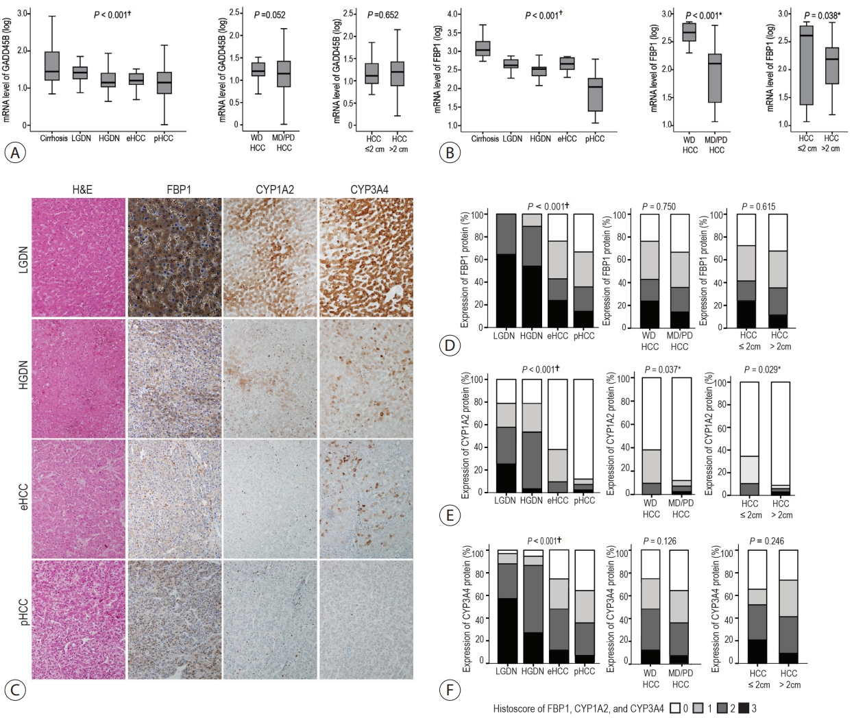
The expression levels of TGF-β signaling genes (TGFB1, TGFBR1, TGFBR2 and SMAD4) gradually increased with the progression of hepatocarcinogenesis, peaking in pHCCs. The expression of early responsive genes of TGF-β (GADD45B, FBP1, CYP1A2 and CYP3A4) gradually decreased, and that of the late TGF-β signatures (TWIST and SNAI1) significantly increased according to the progression of multistep hepatocarcinogenesis. Furthermore, mRNA levels of TWIST and SNAI1 were well correlated with those of stemness markers, with upregulation of TGF-β signaling, whereas FBP1 expression was inversely correlated with that of stemness markers.
Conclusions
The enrichment of the late responsive signatures of TGF-β with induction of stemness is considered to be involved in the progression of the late stage of multistep hepatocarcinogenesis, whereas the early responsive signatures of TGF-β are suggested to have tumor-suppressive roles in precancerous lesions of the early stage of multistep hepatocarcinogenesis.
- Update on Pathologic and Radiologic Diagnosis of Combined Hepatocellular-Cholangiocarcinoma
-
Hyungjin Rhee, Jae Hyon Park, Young Nyun Park
-
J Liver Cancer. 2021;21(1):12-24. Published online March 31, 2021
-
DOI: https://doi.org/10.17998/jlc.21.1.12
-
-
5,729
Views
-
298
Downloads
-
2
Citations
-
Abstract
PDF
- Combined hepatocellular-cholangiocarcinoma (cHCC-CCA) is a malignant primary liver carcinoma characterized by the unequivocal presence of both hepatocytic and cholangiocytic differentiation within the same tumor. Recent research has highlighted that cHCC-CCAs are more heterogeneous than previously expected. In the updated consensus terminology and WHO 2019 classification, “classical type” and “subtypes with stem-cell features” of the WHO 2010 classification are no longer recommended. Instead, it is recommended that the presence and percentages of various histopathologic components and stem-cell features be mentioned in the pathologic report. The new terminology and classification enable the exchange of clearer and more objective information about cHCC-CCAs, facilitating multi-center and multinational research. However, there are limitations to the diagnosis of cHCC-CCA by imaging and biopsy. cHCC-CCAs showing typical imaging findings of HCC could be misdiagnosed as HCC and subjected to inappropriate treatment, if other clinical findings are not sufficiently considered. cHCC-CCAs showing at least one of the CCA-like imaging features or unusual clinical features should be subjected to biopsy. There may be a sampling error for the biopsy diagnosis of cHCC-CCA. An optimized diagnostic algorithm integrating clinical, radiological, and histopathologic information of biopsy is required to resolve these diagnostic pitfalls.
-
Citations
Citations to this article as recorded by  - Differentiation between hepatic angiomyolipoma and hepatocellular carcinoma in individuals who are not at-risk for hepatocellular carcinoma
Sungtae Park, Myeong-Jin Kim, Kyunghwa Han, Jae Hyon Park, Dai Hoon Han, Young Nyun Park, Jaehyo Kim, Hyungjin Rhee
European Journal of Radiology.2023; 166: 110957. CrossRef - The Human TOR Signaling Regulator Is the Key Indicator of Liver Cancer Patients’ Overall Survival: TIPRL/LC3/CD133/CD44 as Potential Biomarkers for Early Liver Cancers
Soo Young Jun, Hyang Ran Yoon, Ji-Yong Yoon, Su-Jin Jeon, Jeong-Ju Lee, Debasish Halder, Jin-Man Kim, Nam-Soon Kim
Cancers.2021; 13(12): 2925. CrossRef
- A Case of Lymphocyte-Rich Hepatocellular Carcinoma in a Patient Who Was Treated for Colon Cancer
-
Jae Won Song, Ho Soo Chun, Jae Seung Lee, Hye Won Lee, Beom Kyung Kim, Seung Up Kim, Jun Yong Park, Sang Hoon Ahn, Young Nyun Park, Dai Hoon Han, Do Young Kim
-
J Liver Cancer. 2021;21(1):69-75. Published online March 31, 2021
-
DOI: https://doi.org/10.17998/jlc.21.1.69
-
-
3,549
Views
-
84
Downloads
-
2
Citations
-
Abstract
PDF
- Hepatocellular carcinoma (HCC) primarily originates in the liver with hepatic differentiation. However, HCCs are not homogenous, and approximately 35% of HCC cases are classified as histopathological variants that present distinct pathologic characteristics. In particular, the lymphocyte-rich variant is the rarest subtype accounting for less than 1% of HCCs, which is not well known to date about molecular features and pathophysiology. Herein, we present a case of a patient who was suspected of metastatic liver cancer and confirmed as lymphocyte-rich HCC pathologically. A 78-year-old woman who underwent a right hemicolectomy for colon cancer was referred to our hospital for a newly detected liver mass. We could not make a decision because of insufficient evidence for diagnosis from imaging studies. After resection, we found that it was a lymphocyte-rich HCC. The pathologic features and prognostic trends of this subtype are also discussed.
-
Citations
Citations to this article as recorded by - Characterization of lymphocyte‐rich hepatocellular carcinoma and the prognostic role of tertiary lymphoid structures
Bokyung Ahn, Hee‐Sung Ahn, Jinho Shin, Eunsung Jun, Eun‐Young Koh, Yeon‐Mi Ryu, Sang‐Yeob Kim, Chang Ohk Sung, Ju Hyun Shim, JeongYeon Hong, Kyunggon Kim, Hyo Jeong Kang
Liver International.2024; 44(5): 1202. CrossRef - Uncommon variants of hepatocellular carcinoma: Not one size fits all
Reetu Kundu, Nalini Gupta, Debajyoti Chatterjee, Ajay Duseja
Diagnostic Cytopathology.2022; 50(1): 28. CrossRef
- Histopathological Variants of Hepatocellular Carcinomas: an Update According to the 5th Edition of the WHO Classification of Digestive System Tumors
-
Haeryoung Kim, Mi Jang, Young Nyun Park
-
J Liver Cancer. 2020;20(1):17-24. Published online March 31, 2020
-
DOI: https://doi.org/10.17998/jlc.20.1.17
-
-
14,202
Views
-
1,003
Downloads
-
27
Citations
-
Abstract
PDF
- Hepatocellular carcinoma (HCC) is heterogeneous in pathogenesis, phenotype and biological behavior. Various histopathological features of HCC had been sporadically described, and with the identification of common molecular alterations of HCC and its genomic landscape over the last decade, morpho-molecular correlation of HCC has become possible. As a result, up to 35% of HCCs can now be classified into histopathological variants, many of which have unique molecular characteristics. This review will provide an introduction to the variously described histopathological variants of HCC in the updated WHO Classification of Digestive System Tumors.
-
Citations
Citations to this article as recorded by - Computational pathology: A survey review and the way forward
Mahdi S. Hosseini, Babak Ehteshami Bejnordi, Vincent Quoc-Huy Trinh, Lyndon Chan, Danial Hasan, Xingwen Li, Stephen Yang, Taehyo Kim, Haochen Zhang, Theodore Wu, Kajanan Chinniah, Sina Maghsoudlou, Ryan Zhang, Jiadai Zhu, Samir Khaki, Andrei Buin, Fatemeh
Journal of Pathology Informatics.2024; 15: 100357. CrossRef - Characterization of lymphocyte‐rich hepatocellular carcinoma and the prognostic role of tertiary lymphoid structures
Bokyung Ahn, Hee‐Sung Ahn, Jinho Shin, Eunsung Jun, Eun‐Young Koh, Yeon‐Mi Ryu, Sang‐Yeob Kim, Chang Ohk Sung, Ju Hyun Shim, JeongYeon Hong, Kyunggon Kim, Hyo Jeong Kang
Liver International.2024; 44(5): 1202. CrossRef - Clinical‐Radiologic Morphology‐Radiomics Model on Gadobenate Dimeglumine‐Enhanced MRI for Identification of Highly Aggressive Hepatocellular Carcinoma: Temporal Validation and Multiscanner Validation
Wanjing Zheng, Xiaodan Chen, Meilian Xiong, Yu Zhang, Yang Song, Dairong Cao
Journal of Magnetic Resonance Imaging.2024;[Epub] CrossRef - Diagnostic Model for Proliferative HCC Using LI‐RADS: Assessing Therapeutic Outcomes in Hepatectomy and TKI‐ICI Combination
Mengtian Lu, Zuyi Yan, Qi Qu, Guodong Zhu, Lei Xu, Maotong Liu, Jifeng Jiang, Chunyan Gu, Ying Chen, Tao Zhang, Xueqin Zhang
Journal of Magnetic Resonance Imaging.2024;[Epub] CrossRef - Low-Baseline PD1+ Granulocytes Predict Responses to Atezolizumab–Bevacizumab in Hepatocellular Carcinoma
Catia Giovannini, Fabrizia Suzzi, Francesco Tovoli, Mariangela Bruccoleri, Mariarosaria Marseglia, Eleonora Alimenti, Francesca Fornari, Massimo Iavarone, Fabio Piscaglia, Laura Gramantieri
Cancers.2023; 15(6): 1661. CrossRef - Non-alcoholic fatty liver disease: the pathologist’s perspective
Wei-Qiang Leow, Anthony Wing-Hung Chan, Paulo Giovanni L. Mendoza, Regina Lo, Kihan Yap, Haeryoung Kim
Clinical and Molecular Hepatology.2023; 29(Suppl): S302. CrossRef - 2022 KLCA-NCC Korea practice guidelines for the management of hepatocellular carcinoma
Journal of Liver Cancer.2023; 23(1): 1. CrossRef - Three-dimensional multifrequency magnetic resonance elastography improves preoperative assessment of proliferative hepatocellular carcinoma
Guixue Liu, Di Ma, Huafeng Wang, Jiahao Zhou, Zhehan Shen, Yuchen Yang, Yongjun Chen, Ingolf Sack, Jing Guo, Ruokun Li, Fuhua Yan
Insights into Imaging.2023;[Epub] CrossRef - Lymphocyte-Rich Hepatocellular Carcinoma with Multiple Lymphadenopathy and Positive Epstein–Barr Virus Encoding Region
Pin-Yi Wang, Yu-Hsuan Kuo, Ming-Jen Sheu, Hsing-Tao Kuo, Wen-Ying Lee, Yu-Ting Kuo, Su-Hung Wang, Zu-Yau Lin
Case Reports in Hepatology.2023; 2023: 1. CrossRef - Genome-Wide Extrachromosomal Circular DNA Profiling of Paired Hepatocellular Carcinoma and Adjacent Liver Tissues
Jianyu Ye, Peixin Huang, Kewei Ma, Zixin Zhao, Ting Hua, Wenjing Zai, Jieliang Chen, Xiutao Fu
Cancers.2023; 15(22): 5309. CrossRef - Proliferative hepatocellular carcinomas in cirrhosis: patient outcomes of LI-RADS category 4/5 and category M
Subin Heo, Hyo Jeong Kang, Sang Hyun Choi, Sehee Kim, Youngeun Yoo, Won-Mook Choi, So Yeon Kim, Seung Soo Lee
European Radiology.2023;[Epub] CrossRef - Cellular heterogeneity and plasticity in liver cancer
Lo-Kong Chan, Yu-Man Tsui, Daniel Wai-Hung Ho, Irene Oi-Lin Ng
Seminars in Cancer Biology.2022; 82: 134. CrossRef - MRI features of histologic subtypes of hepatocellular carcinoma: correlation with histologic, genetic, and molecular biologic classification
Ja Kyung Yoon, Jin-Young Choi, Hyungjin Rhee, Young Nyun Park
European Radiology.2022; 32(8): 5119. CrossRef - Variant Hepatocellular Carcinoma Subtypes According to the 2019 WHO Classification: An Imaging-Focused Review
Liang Meng Loy, Hsien Min Low, Jin-Young Choi, Hyungjin Rhee, Chin Fong Wong, Cher Heng Tan
American Journal of Roentgenology.2022; 219(2): 212. CrossRef - Paraneoplastic syndromes in hepatocellular carcinoma: a review
Yuki Ong, Cheong Wei Terence Huey, Vishalkumar Girishchandra Shelat
Expert Review of Gastroenterology & Hepatology.2022; 16(5): 449. CrossRef - Morphomolecular Classification Update on Hepatocellular Adenoma, Hepatocellular Carcinoma, and Intrahepatic Cholangiocarcinoma
Venkata S. Katabathina, Lokesh Khanna, Venkateswar R. Surabhi, Marta Minervini, Krishna Shanbhogue, Anil K. Dasyam, Srinivasa R. Prasad
RadioGraphics.2022; 42(5): 1338. CrossRef - 2022 KLCA-NCC Korea practice guidelines for the management of hepatocellular carcinoma
Clinical and Molecular Hepatology.2022; 28(4): 583. CrossRef - 2022 KLCA-NCC Korea Practice Guidelines for the Management of Hepatocellular Carcinoma
Korean Journal of Radiology.2022; 23(12): 1126. CrossRef - Histomorphological Subtypes of Hepatocellular Carcinoma and Intrahepatic Cholangiocarcinoma: Review and Update
Yoon Jung Hwang, Haeryoung Kim
AJSP: Reviews and Reports.2022; 27(6): 234. CrossRef - Pediatric Primary Hepatic Tumors: Diagnostic Considerations
Bryony Lucas, Sanjita Ravishankar, Irina Pateva
Diagnostics.2021; 11(2): 333. CrossRef - A Case of Lymphocyte-Rich Hepatocellular Carcinoma in a Patient Who Was Treated for Colon Cancer
Jae Won Song, Ho Soo Chun, Jae Seung Lee, Hye Won Lee, Beom Kyung Kim, Seung Up Kim, Jun Yong Park, Sang Hoon Ahn, Young Nyun Park, Dai Hoon Han, Do Young Kim
Journal of Liver Cancer.2021; 21(1): 69. CrossRef - HCC You Cannot See
Vaishnavi Boppana, Sakshi Sahni, Joseph Glass, Christopher Chang, Denis M McCarthy
Digestive Diseases and Sciences.2021; 66(7): 2185. CrossRef - An update on subtypes of hepatocellular carcinoma: From morphology to molecular
Monika Vyas, Dhanpat Jain
Indian Journal of Pathology and Microbiology.2021; 64(5): 112. CrossRef - HCC: role of pre- and post-treatment tumor biology in driving adverse outcomes and rare responses to therapy
Sandeep Arora, Roberta Catania, Amir Borhani, Natally Horvat, Kathryn Fowler, Carla Harmath
Abdominal Radiology.2021; 46(8): 3686. CrossRef - Gadoxetate-enhanced MRI Features of Proliferative Hepatocellular Carcinoma Are Prognostic after Surgery
Hyo-Jin Kang, Haeryoung Kim, Dong Ho Lee, Bo Yun Hur, Yoon Jung Hwang, Kyung-Suk Suh, Joon Koo Han
Radiology.2021; 300(3): 572. CrossRef - Radiologic Diagnosis of Hepatocellular Carcinoma
Woo Kyoung Jeong
The Korean Journal of Gastroenterology.2021; 78(5): 261. CrossRef - Update on Hepatocellular Carcinoma: a Brief Review from Pathologist Standpoint
Nese Karadag Soylu
Journal of Gastrointestinal Cancer.2020; 51(4): 1176. CrossRef
- Loss of Liver Fatty Acid Binding Protein Expression in Hepatocellular Carcinomas is Associated with a Decreased Recurrence-Free Survival
-
Haeryoung Kim, Hyejung Lee, Young Nyun Park
-
J Liver Cancer. 2015;15(1):30-35. Published online March 31, 2015
-
DOI: https://doi.org/10.17998/jlc.15.1.30
-
-
1,056
Views
-
21
Downloads
-
1
Citation
-
Abstract
PDF
- Background/Aim
s: Loss of liver fatty acid binding protein (LFABP) expression by immunohistochemistry
is a useful marker for the identification of hepatocyte nuclear factor 1α (HNF1α)-
inactivated hepatocellular adenomas; however, the expression status of LFABP in hepatocellular
carcinomas (HCCs) is still unclear. We aimed to investigate the expression status of LFABP
in HCCs and examine the clinicopathological characteristics of LFABP-negative HCCs.
Methods
Immunohistochemical stains LFABP, K19 (mouse monoclonal, Dako, Glostrup, Denmark)
and EpCAM (mouse monoclonal, Calbiochem, Darmstadt, Germany) were performed
on tissue microarray sections from 188 surgically resected HCCs, and the association between
LFABP expression status and the clinicopathological features, survival and “stemness”-related
marker expression status were analyzed.
Results
Loss of LFABP expression was noted in 30 (16%) out of 188 HCCs. LFABP-negative
HCCs were associated with a decreased recurrence-free survival (LFABP-negative: 17.0 ± 4.84
months [95% confidence interval [CI]: 7.5–26.5 months] versus LFABP-positive: 51.0 ± 8.7
months [95% CI: 34.0–68.0 months]; P=0.004). HCCs with LFABP expression loss were more
frequently larger and showed more frequent vascular invasion, although not statistically significant;
and an inverse correlation was seen between LFABP expression and K19 expression
status (P=0.001).
Conclusions
Loss of LFABP expression is seen in HCCs, and is associated with a decreased
recurrence-free survival.
-
Citations
Citations to this article as recorded by - Hepatocellular adenomas: recent updates
Haeryoung Kim, Young Nyun Park
Journal of Pathology and Translational Medicine.2021; 55(3): 171. CrossRef
- A Case of Advanced Hepatocellular Carcinoma which was Supervening with Renal Cell Cancer Cured by Repeated Transarterial Chemoembolization and Sorafenib after Resection
-
Bun Kim, Jae Hoon Min, Seung Up Kim, Jun Yong Park, Kwang Hoon Lee, Do Youn Lee, Jin Sub Choi, Young Deuk Choi, Nam Hoon Cho, Young Nyun Park, Sang Hoon Ahn, Kwang Hyub Han, Chae Yoon Chon, Do Young Kim
-
Journal of the Korean Liver Cancer Study Group. 2012;12(1):51-57. Published online February 28, 2012
-
-
-
Abstract
PDF
- Advanced hepatocellular carcinoma (HCC) is difficult to treat and the survival is poor. Here, we present a patient diagnosed as
advanced HCC (stage IIIa) which was supervening with early renal cell cancer (stage I). The patient was treated with
pre-operational transarterial chemoembolization (TACE) and surgical resection (right hepatectomy, right nephrectomy, and
cholecystectomy). Sorafenib were taken continually after surgery. Multiple recurred HCC nodules in remnant liver were detected
2 months later after surgery. Combined treatment modalities including 4 sessions of TACE, and 12 cycles of 5-flurouracil
(FU)/carboplatin based hepatic arterial infusional chemotherapy (HAIC) induced complete response. After the diagnosis of
advanced HCC, the patient survived 36 months and experienced disease-free status for 19 months.
- A Case of Early Recurred Hepatocellular Carcinoma with Initial Expectation of Good outcome by Surgical Resection
-
Jung Min Lee, Jun Yong Park, Do Young Kim, Kyung Sik Kim, Young Nyun Park, Myeong-Jin Kim, Chae Yoon Chon, Kwang-Hyub Han
-
Journal of the Korean Liver Cancer Study Group. 2009;9(1):41-44. Published online June 30, 2009
-
-
-
Abstract
PDF
- Hepatocellular carcinoma (HCC) is one of the cancers with poor prognosis as HCC develops on base of cirrhosis in
majority cases, which requires multidisciplinary approach. If feasible, however, surgical resection is the choice of treatment,
and many previous studies and guidelines offered appropriate indications for surgical resection; firstly, preservation of liver
function should be confirmed with traditional Child-Pugh classification or more recently with Indocyanine Green retention test
or absence of portal hypertension. Secondly, several variables about the size, number, and vascular invasion of tumor should
be taken into consideration. It is suggested that to lessen the risk of recurrence gross vascular invasion be absent and the
number of tumor be single. Regarding the size of tumor, although risk of dissemination increases with size, some tumors
may grow as a single mass and thus the size of tumor is not a clear-cut limiting factor. Based on above suggestions, we
herein offer our experience of a patient with initial hopeful expectation of good outcome with surgical resection, but who
eventually turned out to result in disseminated tumor recurrence. Further study, maybe regarding a combination of adjuvant
or neoadjuvant transarterial chemoembolization/chemotherapy or radiotherapy, is necessary on how to manage such patient.
- A Case of Hepatocellular Carcinoma in a Pregnant Patient in Twenties
-
Kang Kook Choi, Young Ju Hong, Sae Byeol Choi, Nam Joon Yi, Shin Hwang, Young Nyun Park, Jin Sub Choi, Kyung Suk Suh, Chae Yoon Chon, Kyung Sik Kim
-
Journal of the Korean Liver Cancer Study Group. 2009;9(1):76-81. Published online June 30, 2009
-
-
-
Abstract
PDF
- Hepatocellular carcinoma (HCC) in pregnancy is very rare. The cirrhosis which is present in the majority of patients with
HCC induces infertility. The diagnostic methods and treatment modalities in HCC during pregnancy are different from those
of usual types of other HCC. A 26-year-old, 32th-gestational-week pregnant female was sent to our hospital because of
abnormal liver function test. A 1.5cm sized mass was identified in segment 6 of liver which was compatible to AJCC stage
I. She did not have any other medical history except Hepatitis B Virus carrier and the HBs Ag of her mother also was
positive. At the 40th gestational week, the female baby was delivered uneventfully. And then she underwent the transarterial
chemoembolization (TACE) following the Rt. Hemihepatectomy. Since she underwent a surgical resection, the tumors have
been recurred in the remnant liver only. Whenever the tumors were founded, the aggressive surgical approaches were
performed including 3 times of hepatic resection with TACE or TACI. She is still alive with good general condition and
normal liver function for 9 years since the first diagnosis was made. Therefore an extremely rare case of hepatocellular
carcinoma in pregnancy is treated successfully because of aggressive therapies.
- A Case of Curative Resection of Advanced Hepatocellular Carcinoma After Localized Concurrent Chemo-Radiation Therapy
-
Wonseok Kang, Ki Tae Yoon, Jun Yong Park, Do Young Kim, Sang Hoon Ahn, Chae Yoon Chon, Kyung Sik Kim, Young Nyun Park, Jin Sil Seong, Kwang Hyub Han
-
Journal of the Korean Liver Cancer Study Group. 2008;8(1):98-101. Published online June 30, 2008
-
-
-
Abstract
PDF
- Most patients with advanced hepatocellular carcinoma (HCC) are not suitable candidates for surgical treatment
at the time of diagnosis because of poor liver function, extensive tumor involvement of the liver, vascular
involvement, and/or intra/extrahepatic metastasis. We attempted localized concurrent chemo-radiation therapy
(CCRT) followed by hepatic arterial infusion chemotherapy (HAIC) in patients having locally advanced HCC with
vascular involvement and preserved hepatic function. We report a case of locally advanced HCC patient who
became surgically resectable by downstaging after localized CCRT followed by HAIC. Localized CCRT was
performed with a total radiation dose of 4,500 cGy (180 cGy × 25 times) and hepatic arterial infusion of
5-fluorouracil (5-FU, 500 mg/day) via implantable port system during the first and the last weeks of the
radiotherapy. Following localized CCRT, the patient was scheduled to receive HAIC with 5-FU (500 mg/m2 for
5 hours, days 1~3) and cisplatin (60 mg/m2 for 2 hours, day 2) every 4 weeks. Marked contraction of HCC was
noted on follow up computerized tomography (CT) and positron emission tomography (PET) after localized CCRT
and HAIC, and subsequently surgical resection with curative aim was performed. The patient is in complete
remission status without recurrence to date.
- Primary hepatic carcinoma with mixed features
-
Young Nyun Park
-
Journal of the Korean Liver Cancer Study Group. 2007;7(1):13-15. Published online June 30, 2007
-
-
-
PDF
- A Case of Focal Nodular Hyperplasia-like Nodules in Cirrhosis
-
Young Joon Yoon, Ki Tae Yoon, Jun Yong Park, Hyun Woong Lee, Hwa Sook Kim, Jae Kyung Kim, Young Nyun Park, Kwang-Hyub Han, Chae Yoon Chon, Young Myung Moon, Mi-Suk Park, Sang Hoon Ahn
-
Journal of the Korean Liver Cancer Study Group. 2007;7(1):41-44. Published online June 30, 2007
-
-
-
Abstract
PDF
- Focal nodular hyperplasia (FNH) usually occurs in non-cirrhotic livers and was defined as a nodule composed
of benign appearing hepatocytes occurring in a liver that is otherwise histologically normal or nearly normal.
However, due to improvements in imaging techniques and pathological evaluation of explant livers, a focal lesion
that is very similar to the classic form of focal nodular hyperplasia that occurs in cirrhotic liver has been
described by several reports. Therefore, the term FNH-like nodules has been proposed. In this report, we report
a case of focal nodular hyperplasia-like nodules in cirrhosis. A 59 year old woman with known hepatitis B virus
infection visited our institution for routine check up. She was diagnosed as having liver cirrhosis and 3.5 cm sized
liver mass on abdomen ultrasonography (US). Because tumor marker was negative and US findings are not
compatible with hepatocellular carcinoma, other imaging modalities were performed. Magnetic resonance imaging
(MRI) documented a 3.5 cm sized hypervascular nodule with internal aberrant vascular structure and multiple
small sized nodules in remaining liver. Needle biopsy was targeted to the liver main mass. Microscopic finding
revealed FNH-like nodule and underlying liver cirrhosis.
- Two cases of Vessel invasion of Hepatocellular carcinoma
-
Sae Byeol Choi, Jae Yeon Seok, Kyung Sik Kim, Young Nyun Park, Kwang-Hyub Han, Jong Tae Lee
-
Journal of the Korean Liver Cancer Study Group. 2007;7(1):71-76. Published online June 30, 2007
-
-
-
Abstract
PDF
- Despite growing information on the clinical behavior of hepatocellular carcinoma(HCC), the histologic features
associated with survival are not well characterized. Several different staging systems are suggested for use in
predicting the prognosis of HCC. American Joint Committee on Cancer/International Union Against Cancer
Staging System (AJCC/UICC) 6th edition divided T stages according to vessel invasion, T1 without microvessel
invasion, T2 showing microvessel invasion and T3 showing major vessel invasion. The vessel invasion is
generally considered a poor prognostic factor for HCC. Our report of the two patients with HCC run along similar
terms. The patient diagnosed HCC with microvessel invasion underwent left lateral sectionectomy. Although the
presence of microvessel invasion was found, this patient has survived without any recurrence for over 5 years
now. The other patient underwent S8 segmentectomy and lived 10 years disease-free. After 10 years, although
an intrahepatic recurred HCC successfully treated with local therapy, the recurred and newly developed multiple
lesions were found again leading to a decision to perform operation. The HCC invaded into the portal vein and
constituted portal vein thrombosis. The patient expired after 3 months postoperatively due to intrahepatic
dissemination of the tumor. Therefore the impact of the vascular invasion on long-term survivors remains to be
determined.
- A Case of Advanced Hepatocellular Carcinoma with Inferior Vena Caval Invasion, Resected with a Curative Aim after Concurrent Chemo-Radiation Therapy
-
Seung Up Kim, Kwang Hyub Han, Jin Sil Sung, Do Young Kim, Sang Hoon Ahn, Gyeong Sig Kim, Young Nyun Park, Chae Yoon Chon
-
Journal of the Korean Liver Cancer Study Group. 2007;7(1):77-81. Published online June 30, 2007
-
-
-
Abstract
PDF
- Hepatocellular carcinoma (HCC) with tumor thrombus in inferior vena cava (IVC) is difficult to treat. Therefore,
there are no specific treatment modalities for such case. Here, we present a patient diagnosed as hepatocellular
carcinoma with tumor thrombus in inferior vena cava (stage IVa). The patient was treated with concurrent
chemo-radiation therapy (CCRT) for 5 weeks. After that, tumor size was markedly decreased, and 9th courses of
additional intra-arterial chemotherapy were performed. Follow-up positron emission tomography- computed
tomography (PET-CT) showed shrinked hepatocellular carcinoma and right lobe, disappearance of IVC tumor
thrombus, decreased size of right hepatic vein thrombus and a faint uptake at gallbladder. Residual malignancy
could not be excluded. So, right hepatic lobectomy with a curative aim was performed and its result was
successful.
- Well-differentiated Samll Hepatocellular Carcinoma, Early Hepatocellular Carcinoma and Dysplastic Nodule
-
Young Nyun Park
-
Journal of the Korean Liver Cancer Study Group. 2004;4(1):7-11. Published online June 30, 2004
-
-
-
PDF
- Pathologic Findings of Variants and Uncommon Types of Primary Liver Cancers
-
Young Nyun Park
-
Journal of the Korean Liver Cancer Study Group. 2002;2(1):27-35. Published online July 31, 2002
-
-
-
PDF
- A Case of Hepatocellular Carcinoma with Dramatic Therapeutic Response in Stage Ⅳa (T4N0M0)
-
Hye Won Chung, Jae Youn Cheong, Kun Hoon Song, Kwang Hyub Han, Young Myoung Moon, Byoung Ro Kim, Jin Sil Seong, Myoung Jin Kim, Do Yun Lee, Young Nyun Park
-
Journal of the Korean Liver Cancer Study Group. 2001;1(1):89-92. Published online June 30, 2001
-
-
-
Abstract
PDF
- A 52 year-old-female patient was admitted due to right upper quadrant abdominal pain and right shoulder pain for 1 month. She had no history of previous medical problem before admission. At admission, physical findings revealed that she had 2 FB palpable hepatomegaly and others were not remarkable. Laboratory findings revealed that HBsAg and HBeAG were positive enhanced at arterial phase involving entire right lobe and S4, and thrombus at main portal vein. Clinically she was diagnosed advanced hepatocellular carcinoma(HCC) and TNM stage was T4N0M0, stage Ⅳa. She was treated with transarterial chemoembolization(TACE) for 2 cycles and radiation therapy. After TACE and radiation therapy, the tumor size decreased and the main portal vein thrombus was disappeared. Then, she underwent extended right lobectomy of the liver. Pathology of operative specimen revealed well-encapsulated HCC at Edmonsons grade Ⅱ, near total necrosis. Normal liver tissue showed sinusoidal congestion, mild periportal fibrosis, and veno-occlusive disease. Two years after the operation, there had been no sign of recurrence and follow-up CT scan showed sign of recurrence at S2. He underwent percutaneous Holmium injection 3 times and TACE 4 times, and so far he survived for 6 years and 10 months. He is currently under observation at out patient department.
|
 E-submission


 Follow JLC on Twitter